Presenting two case reports of urachal anomalies.
Submitted by Dr MGK Murthy, Dr GA Prasad
Case 1(MRI)
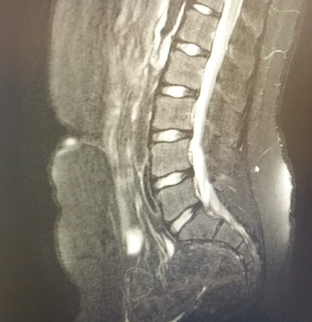
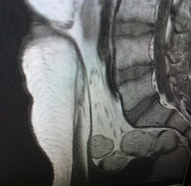
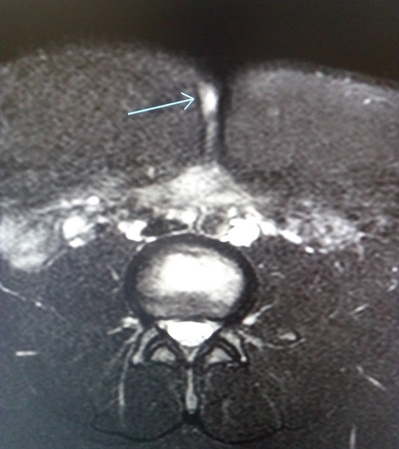
14 yrs boy with h/o periumbilical pain, swelling & discharge with periumbilical sinus in USG presents for MRI which show- An ill defined irregular subtle fluid signal intensity focus suggested in the infraumbilical region with a long thin linear low signal intensity on all pulse sequences properitoneal track identified extending to the superior aspect of urinary bladder with no definite fluid contents/ bladder diverticulum/secondary tracks/intraperitoneal extension/ presence of air /air fluid levels – likely represents urachal anomaly in the form urachal umbilical sinus track with no significant cyst formation /vesicourachal diverticulum.
Case 2 ( CT scan)
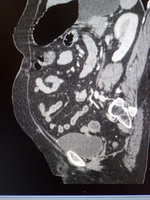
58 yrs female presents with pain abdomen for CT scan shows thin cord like nonenhancing hypodense track from urinary bladder dome upto the umbilicus with small speck of calcification at the bladder end with no sinus or cyst or bladder diverticulum suggesting urachus without any complications.
Urachal anomalies result from abnormal persistence of embryologic communication between umbilicus & urinary bladder. During normal gestational development, the urachus involutes and its lumen is obliterated, becoming the median umbilical ligament. Urachal anomalies are – Patent urachus or urachal fistula– patent connection between umbilicus & urinary bladder. Umbilical urachal sinus – blind focal dilatation at umbilical end. Vesicourachal diverticulum – focal outpouching at bladder end. Urachal cysts – midline cysts near the bladder end usually.
Common complications of urachal remnants are infection & tumors of urachus. Treatment – surgical excision of urachal remnant.
Famous Radiology Blog http://bit.ly/1MM2hKr TeleRad Providers at http://bit.ly/1NgppuI Mail us at sales@teleradproviders.com
from Imaging via a.sfakia on Inoreader http://bit.ly/2Sftjib


No comments:
Post a Comment
Note: Only a member of this blog may post a comment.